The ability to stain and detect intracellular molecules opens the door to identify distinct cell subsets as well as further characterize cell populations. Our resources, tools and products help you save time and effort and increase your efficiency when designing your flow cytometry experiments. In this blog entry, we are going to show you five key points to save time and efforts while you are designing your experiments of flow cytometry.
- Determine the non-nuclear intracellular staining
- Considerations to perform Nuclear staining
- Intracellular staining buffer selection
- Flow cytometry for studying the cell signaling pathways
- Add to your flow cytometry workflow the compensation beads UltraComp™ eBeads
Determine the non-nuclear intracellular staining
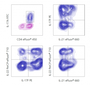
Staining protein inside the cell requires fixation to crosslink proteins and stabilize the cell membrane followed by permeabilization to allow antibodies access to intracellular antigens. Intracellular fixation and permeabilization buffers are designed for optimal detection of cytoplasmic proteins and secreted proteins residing in organelles and vesicles, such as cytokines and chemokines.

Because secreted proteins are generally expressed at low levels in resting cells, expression must be inducd and secretion blocked to allow for detection by flow cytometry. While PMA (Phorbol 12-myristate 13-acetate) and Ionomycin (calcium ionophore) are often used in combination to induce cytokine production, for more specific stimulation or cell-type activation, agonistic antibodies against cell receptors, such as CD3 and CD28 for T lymphocytes, are a great option.
Once the proteins are expressed, it is necessary to block the secretory pathway to allow for accumulation of the proteins of interest. This is often achieved with Brefeldin A, which blocks transport at the endoplasmic reticulum, or with Monensin which blocks transport at the Golgi apparatus. Check eBioscience’s catalog for the Cell Stimulation Cocktail:

Due these characteristics, we can find many solutions in eBioscience’s catalog to take advantage of your project.
Considerations to perform Nuclear staining
Transcription factors are DNA-binding proteins that regulate gene expression by modulating the synthesis of messenger RNA. A greater understanding of the expression and regulation of transcription factor activity in immune cells may reveal novel therapeutic opportunities.
Intracellular staining buffer selection
When performing intracellular staining followed by flow cytometric analysis, the selection of fixation and permeabilization buffer systems has a significant impact on the quality and accuracy of the data.
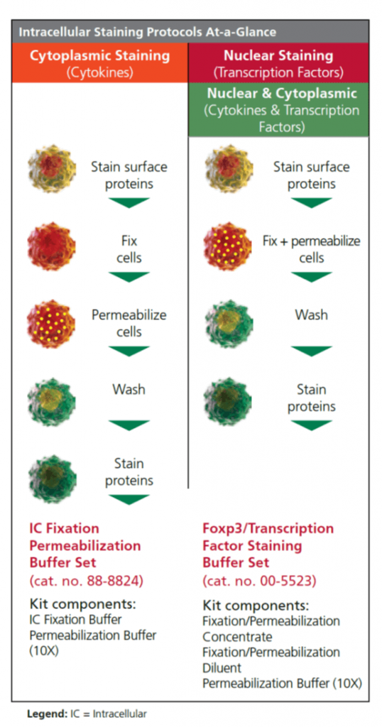
It is important to consider the location of the target proteins within the cell in order to slect the appropriate buffer. For example, to obtain optimal staining of a transcription factor, the Foxp3/Transcription Factor Staining Buffer Set is recommended; however, secreted proteins such as cytokines and chemokines work best with the Intracellular Fixation and Permeabilization Buffer Set.
When staining of proteins that localize to different regions of a cell, the correct buffer choice becomes more challenging. Each antibody should be optimized independently to validate the staining pattern. The chart below provides general rules to follow when choosing the appropriate buffer system.
Flow cytometry for studying the cell signaling pathways
Flow cytometric analysis of phosphorylated proteins using phospho-specific antibodies provides reseaarchers an effective way to elucidate signaling cascades in individual cells. eBioscience offers a portfolio of phospho-specific antibodies for flow cytometry that has been validated in a variety of experiments to give scientists the confidence that these antibodies will perform robustly and reliably for:
- Pathway-specific testing.
- Cell type-specific testing.
- Application testing: Western blot, ELISA, and Immunochemistry.
- Performance in different intracellular fixation/permeabilization buffers.
- Mouse and human cross-reactivity testing.

LEFT: Western blotting of reduced lysates from Jurkat cells unstimulated left lane), stimulated with PMA and Ionomycin middle lane), or stimulated with PMA and Ionomycin in the presence of the MEK1/2 inhibitor, PD98059 right lane) using Anti-Human/Mouse phospho-ERK1/2 T202/Y204) Purified. RIGHT: Intracellular staining of Jurkat cells that were unstimulated black histogram), stimulated with PMA and Ionomycin green histogram), or stimulated with PMA and Ionomycin in the presence of the MEK1/2 inhibitor, PD98059 red histogram) with Anti-human/Mouse phospho-ERK1/2 T202/Y204 APC
Add to your flow cytometry workflow the compensation beads UltraComp™ eBeads

Staining of UltraComp eBeads with 13 different eFluor 450-conjugated monoclonal antibodies including one of each subclass commonly used in flow cytometry. Beads were stained with 0.25 ug of each antibody and analyzed by flow cytometry. Each histogram represents one staining antibody clone and isotype indicated at right)
UltraComp eBeads™ react with antibodies of mouse, rat and hamster origin, and are immunoglobulin light chain-independent. They are designed for use in compensation with all fluorochromes excited by ultraviolet (355 nm), violet (405 nm), blue (488 nm), green (532 nm), yellow-green (561 nm), and red (633-640 nm) lasers. The beads are spherical particles that can be stained with individual fluorochrome-conjugated antibodies for use as single-color compensation controls.
Each drop of beads contains two populations: a positive population that will capture any mouse, rat or hamster antibody and a negative population that will not react with antibody. When a fluorochrome-conjugated antibody is added to the beads, both positive and negative populations result. This bimodal distribution can be used for single-color compensation controls in multicolor flow cytometry experiments.
¿Tienes dudas?
Si no te queda claro del todo cómo funciona esta tecnología, o quieres que te ayudemos a configurar tu ensayo, nuestro departamento técnico de especialistas, con amplia trayectoria en investigación (todos PhD), te pueden echar una mano: por mail (tecnic@labclinics.com), por tlf +34.934464700 o de forma presencial. Contáctanos y estaremos encantados de poder ayudarte!


Leave a reply