
Many scientists working in oncology focus on cancer stem cells (CSCs). Targeting these cells specifically could improve cancer therapy significantly. Because CSCs rely heavily on their environment, three-dimensional cultivation offers the unique opportunity to better understand this interaction. We talked to two scientists working in this field of cancer research.
- Who are the Cancer Stem Cells?
- The importance of a tridimensional structure
- Researchers need 3D cell cultures to better understand the world of cancer stem cells
- Do it yourself: Establishing 3D tumors cell cultures
- Main characteristics of the Promocell Primary Cancer Stem Cell System
- References
Who are the Cancer Stem Cells?
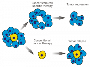 Cancer stem cells (CSCs) are cancer cells that possess characteristics associated with normal stem cells, specifically the ability to give rise to all cell types found in a particular cancer sample. Such cells are hypothesized to persist in tumors as a distinct population and cause relapse and metastasis by giving rise to new tumors.
The first conclusive evidence for CSCs came in 1997 in Nature Medicine. Bonnet and Dick¹ isolated a subpopulation of leukemia cells that expressed surface marker CD34, but not CD38. The authors established that the CD34+/CD38− subpopulation is capable of initiating tumors in NOD/SCID mice that were histologically similar to the donor.
Get more information regarding this complex group of cancer cells, following this link.
The importance of a tridimensional structure
In the human body, two-dimensional structures do not exist. Cells are never present as single-cell entities, but are always surrounded by other cells, forming three-dimensional tissues and organs. In addition, they are embedded in a complex non-cellular structure known as the extracellular matrix. When considering this physiology, it is perhaps surprising that until the mid-90s, two-dimensional models were used in cell culture to analyze tumor cells. In 1996, Reynolds and colleagues showed that undifferentiated neural cells could be grown in suspension as neurospheres. Since then, numerous three-dimensional tumorsphere assays have been established, and tumorsphere cultivation is now widely used to study cancer cells, as well as to screen potential anticancer agents.
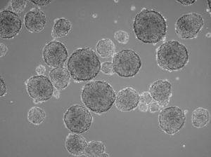
Example of a tumorsphere culture of HT1080 fibrosarcoma cells: A robust tumorsphere formation can be observed in the HT1080 cancer cells cultured with the 3D 3D Tumorsphere Medium XF (picture from Promocell after 10 passages)
Insights from such experiments are more translatable to conditions in patients than results from the conventional two-dimensional monolayer approach. Cells in three-dimensional culture also differ considerably in morphology, proliferation rate, differentiation, apoptosis and gene expression from adherent cells. In fact, cells that are capable of shaping tumorspheres build a “home” for cancer cells, and, in particular, for cancer stem cells (CSCs).
Researchers needs 3D cell cultures to better understand the world of cancer stem cells
About twenty years ago, cancer stem cells were first described by researchers, this field of research has grown, and CSCs have been identified in various types of solid tumors, including those in the brain, breast, colon, head and neck, liver and lung (Lee et al.²). Now CSCs are considered to be the primary cause of cancer recurrence, as they are highly resistant to current chemotherapy and radiotherapy regimens. This is why they represent a key target for successful cancer treatment and their elimination could lead to permanent remissions.
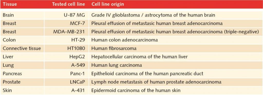
3D cell culture tested cell lines: The table shows cell types successfully tested for serial passages with the 3D Tumorsphere Medium XF
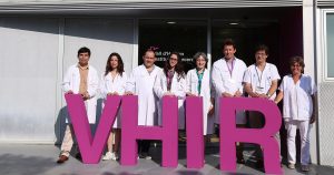
Dr. Matilde E. Lleonart and her team from the Vall d’Hebron Research Institute in Barcelona grow tumorspheres from a single CSC, with the aim of finding new molecular markers
The greatest obstacle to studying cancer stem cells is the isolation and culture of sufficient numbers of cells. Characterization is also difficult, as CSCs are a highly heterogeneous cell population lacking specific markers. Solving these issues is the aim of the team led by Dr. Matilde E. Lleonart at Vall d’Hebron Research Institute (VHIR) in Barcelona, Spain.“At the moment, there are no specific markers for CSCs, so scientists routinely use the general characteristics that apply to all stem cells. We want to identify unique features that will help to better characterize CSCs,”“, explains the Dr. Lleonart.
“Our work involves creating in vitro models that resemble the in vivo situation as closely as possible. This allows us to study the molecular and phenotypical changes of CSCs under different conditions.” The team is using three-dimensional tumorsphere cultivation to enrich CSCs from bulk cancer cells. They are isolating and studying stem cells from breast, lung, and head and neck cancer, to better understand the cells’ implications in tumor biology.
Do it yourself: Establishing 3D tumor cell cultures
Three-dimensional models also provide valuable insights into the interactions between host and tumor. Researchers can better investigate the tumor microenvironment, which plays a significant role in cancer progression (Nath S. et al.³). Tumorsphere cultivation is based on culturing cancer cells onto an ultralow attachment surface in serum-free media (Lee CH et al.²). Researchers can use this technique mainly for two purposes, namely either for enriching cancer stem cells or for isolating primary cells from human tumor samples and maintain the cancer cell heterogeneity.
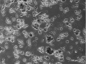
Culture of Primary renal cell carcinoma cells. Cancer cells showing a tumorsphere-forming growth pattern on day 13 of culture.
Both approaches use the advantage of the 3D structures to build up a more physiological microenvironment.
Whereas Dr. Lleonart pursues the first goal using the PromoCell 3D Tumorsphere Medium XFJulia Schnappinger, a postgraduate researcher at the Immunoanalytics Core Facility of Helmholtz Zentrum in Munich (German Research Center for Environmental Health) wants to mantain the different cancer cell populations including the CSCs with the Primary Cancer Culture System. La Dra. Schnappinger explains: “For our studies, it is important that the lymphocytes maintain the same properties in vitro they have in vivo“. When cultured, TILs tend to alter their phenotypical characteristics, as they are no longer conditioned by the cancer cells. Yet tumorspheres perfectly resemble the tumor environment, thus maintaining the original tumor properties while offering the best surroundings for the TILs.”
Main characteristics of the Promocell Primary Cancer Stem Cell System
The PromoCell Primary Cancer Culture System offers a defined and animal component-free system for the isolation and culture of human primary tumor cells. Although the use of three-dimensional cultures has significant advantages, working with tumorspheres can be difficult when using primary cells or fresh tissues. “Compared to monolayer cultures, it is sometimes difficult to find the right time to split the spheroids. You need a lot of experience. If you do this too late, the cells will start to die.” remarks Schnappinger. Similar observations are reported by Lleonart: “It is more difficult to establish 3D cultures than monolayers, although it is somewhat easier when you are working with cell lines. When working with primary cells, the size of the biopsies is crucial.“.
 Both scientists obtained significantly better results with PromoCell 3D Tumorsphere Medium XF and the primary culture system for cancer. “Phenotypically, we could not observe any difference between cells grown in homemade media and cells grown in PromoCell Media. However, when we looked at stem cell gene expression, the purity of the CSC population was higher when working with 3D Tumorsphere Medium XF”, explains Lleonart.
In conclusion, three-dimensional-tumorspheres build a “home” for cancer cells that are enriching and expanding tumor cells. Using 3D cultivation gives researchers new insights into cancer biology, and tumorspheres provide important resources for cancer studies. In a world where personalized therapy is becoming a realistic option for many cancer patients, tumorspheres could be precious resources for evaluating the efficacy of treatments prior to their use in patients. By investigating cancer cells in a biologically relevant in vitro model, researchers will better understand cancer, which ultimately could lead to new therapies.
References
- Bonnet D and Dick JE. (1997) Human acute myeloid leukemia is organized as a hierarchy that originates from a primitive hematopoietic cell. Nat Med 3:730-7. Entrez PubMed 9212098
- Lee CH (2016) Tumorsphere as an effective in vitro platform for screening anti-cancer stem cell drugs. Oncotarget 7:1215-1226
- Nath S (2016) Three-dimensional culture systems in cancer research: focus on tumor spheroid model. Pharmacol Ther 163:94-108
You have doubts?
If it is not entirely clear to you how this technology works, or you want us to help you set up your essay, our technical department of specialists, with extensive experience in research (all PhD), can lend you a hand: by mail (tecnic@labclinics.com), by telephone +34.934464700 or in person. Contact us and we will be happy to help you!


Leave a reply