
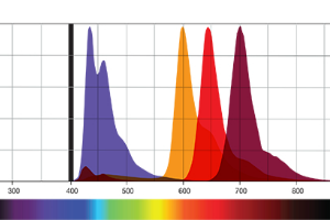
The eBioscience™ Super Bright dyes are a line of bright fluorochromes, based on a polymer and its tandems, that are excited by the violet laser (405 nm). All Super Bright formats are named for their emission wavelength.
- Are they compatible with other reagents?
- SUPER BRIGHT 436
- SUPER BRIGHT 600
- SUPER BRIGHT 645
- SUPER BRIGHT 702
- SUPER BRIGHT STAINING BUFFER
These dyes are optimized for use in flow cytometry and may allow for better discrimination of dim populations due to their brightness.
Are they compatible with other reagents?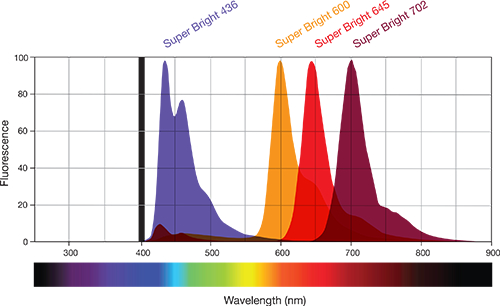
The answer is YES.
Certain dyes may display less nonspecific interaction with other polymer dyes as compared to similar competitor reagents. The Super Bright polymer dyes are fully compatible with other commonly used fluorescent molecules, eBioscience buffers and fixatives, and UltraComp eBeads™ microspheres.
These features, combined with our broad portfolio of biological content, easily enable dye selection for optimized flow cytometry multicolor antibody panel design and allow you to expand the utility of your violet laser.
Here’s a brief review:
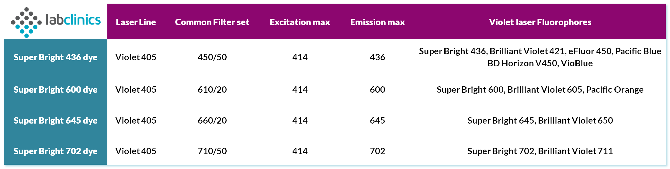
SUPER BRIGHT 436
Super Bright 436 has an excitation maximum of 414 nm and an emission peak of 436 nm. We recommend using a 450/50 bandpass filter or equivalent, similar to eFluor™ 450. This polymer dye is significantly brighter than eFluor 450, and is an alternative for Brilliant Violet™ 421 with similar resolution of positive and negative populations (Figure 2). Stability studies indicate that Super Bright 436 exhibits a minimal loss of fluorescence when cells are exposed to formaldehyde fixative for up to three days, or overnight to ambient light.
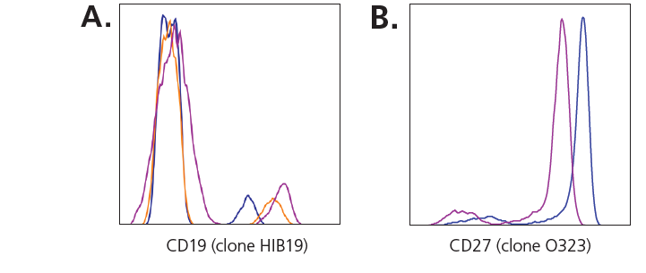
Fluorescence intensity comparison
(A) Human peripheral blood cells were stained with Anti-CD19 (clone HIB19) conjugated to either Super Bright 436 (purple histogram), eFluor 450 (blue histogram), or Brilliant Violet 421 (orange histogram) using the manufacturer’s recommended volume per test. (B) Anti-human peripheral blood cells were stained with Anti-CD27 (clone O323) conjugated to either Super Bright 436 (purple histogram) or Brilliant Violet 421 (blue histogram)
SUPER BRIGHT 600
Super Bright 600 is a tandem dye consisting of Super Bright 436 and an acceptor dye that emits at 600 nm. It can be detected using a 610/20 bandpass filter or equivalent, similar to eVolve™ 605. This tandem polymer dye is comparable in brightness to Brilliant Violet™ 605 and is brighter than eVolve 605. Super Bright 600 is stable for up to three days when stored in a formaldehyde fixative solution.
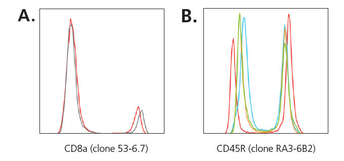
Staining performance and post-fixation stability
(A) Direct comparison of mouse splenocytes stained with Anti-CD8a (clone 53-6.7) conjugated to either Super Bright 600 (red histogram) or Brilliant Violet 605 (gray histogram), at the same concentration of antibody.
(B) Mouse splenocytes were stained with Anti-CD45 (clone RA3-6B2) Super Bright 600 and either left unfixed (red histogram), or were fixed in IC Fixation buffer for 30 minutes (blue histogram), 24 hours (orange histogram), or three days (green histogram)
SUPER BRIGHT 645
Invitrogen eBioscience Super Bright 645 dye is tandem polymer dye, consisting of violet (405 nm) laser–excitable Super Bright 436 and an acceptor dye that emits fluorescence signal at 645 nm. The technology behind the Super Bright polymer dye family makes them useful for identifying low-abundance markers on a cell surface in flow cytometry because they emit fluorescence with high efficacy.
An alternative to Brilliant Violet 650 conjugates, antibody conjugates of Super Bright 600 provide increased resolution of positive and negative populations, but with less background and less interfering dye–dye interaction than Brilliant Violet 650 conjugates.
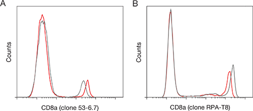
Fluorescence intensity comparison with Super Bright 645 dye (A) Mouse splenocytes stained with CD8a antibody (clone 53-6.7) conjugated to Super Bright 645 dye (red histogram) or Brilliant Violet 650 dye (gray histogram), at the same concentration of antibody. (B) Human peripheral blood cells stained with CD8a antibody (clone RPA-T8) conjugated to Super Bright 645 dye (red histogram) or Brilliant Violet 650 dye (gray histogram), using the same concentration of antibody.
SUPER BRIGHT 702
Invitrogen eBioscience Super Bright 702 dye is tandem polymer dye, consisting of violet (405 nm) laser–excitable Super Bright 436 and an acceptor dye that emits fluorescence signal at 702 nm. The technology behind the Super Bright polymer dye family makes them useful for identifying low-abundance markers on a cell surface in flow cytometry because they emit fluorescence with high efficacy.
An alternative to Brilliant Violet 711 conjugates, antibody conjugates of Super Bright 702 provide increased resolution of positive and negative populations, but with less background and less interfering dye–dye interaction than Brilliant Violet 711 conjugates.

Fluorescence intensity comparison with Super Bright 702 dye (A) Mouse splenocytes stained with CD4 antibody (clone GK1.5) conjugated to Super Bright 702 dye (red histogram) or Brilliant Violet 711 dye (gray histogram), at the same concentration of antibody. (B) Human peripheral blood cells stained with CD19 antibody (clone HIB19) conjugated to Super Bright 702 dye (red histogram) or Brilliant Violet 711 dye (gray histogram), using the same concentration of antibody
SUPER BRIGHT STAINING BUFFER
Super Bright dyes can be used in flow cytometric applications similarly to traditional fluorophores. However, if multiple Super Bright-conjugated antibodies are combined in the same panel, the use of Super Bright Staining Buffer is recommended to minimize any nonspecific interaction that may occur between these polymer-based dyes.
No special buffer is required when using a single Super Bright-conjugated antibody within a panel.
When using Super Bright dyes in combination with other polymer dyes, such as Brilliant Violet dyes, the Super Bright Staining Buffer can still be used to prevent dye–dye interaction. Super Bright Staining Buffer is formulated at 5 µL/test, making it convenient for use when preparing cocktails.
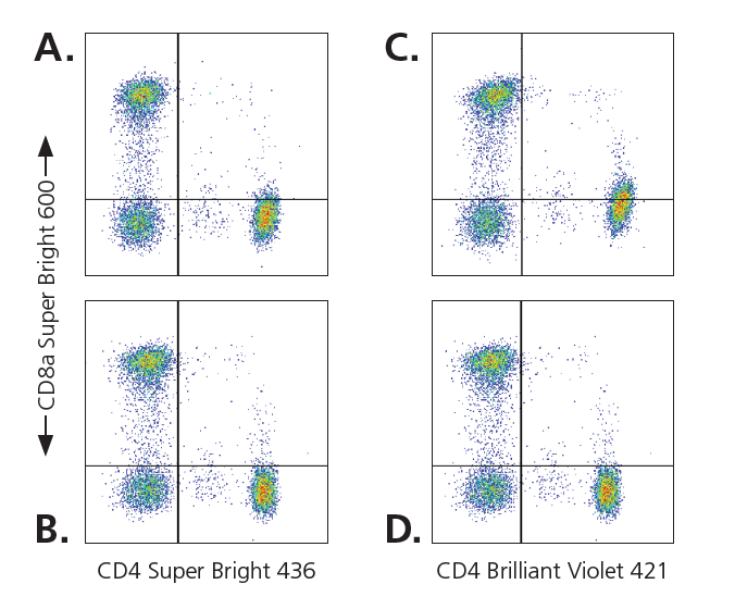
Super Bright Staining Buffer minimizes non-specific interactions
Human peripheral blood cells were stained with Anti-CD8 (clone RPA-T8) Super Bright 600 and Anti-CD4 (clone SK3) conjugated to Super Bright 436 (A and B) or Brilliant Violet 421 (C and D). Cells were stained in the presence of Flow Staining Buffer only (A and C) or Super Bright Staining Buffer was added to cells prior to addition of antibodies (B and D)
ANY QUESTIONS?
If you have some questions about how this technology works, or need any help to set up your assay, our experienced Tech Support Team (all are PhD) will be happy to help you by mail (tecnic@labclinics.com), phone (+34.934464700) or we can visit you. Let’s talk!

Leave a reply